Scaffold-Based Bladder Cancer Modeling Service
Three-dimensional (3D) cell scaffolds are designed to mimic the natural extracellular matrix (ECM) in bladder cancer tissues and organs, supporting cell growth and organization in a three-dimensional environment. Alfa Cytology offers bladder cancer modeling services based on various material scaffolds.
3D Cellular Scaffold Models for Bladder Cancer
3D cell culture systems are becoming increasingly popular in bladder cancer research, tissue engineering, drug discovery, and drug resistance studies. In comparison to conventional two-dimensional (2D) cell culture models, the 3D cellular scaffold model presents several notable benefits. It creates a microenvironment that more accurately reflects physiological conditions by enabling cells to grow in 3D, departing from the limitations of flat surfaces. Consequently, this closer approximation to the native tissue architecture allows for enhanced cell-cell and cell-matrix interactions, closely resembling those observed in vivo.
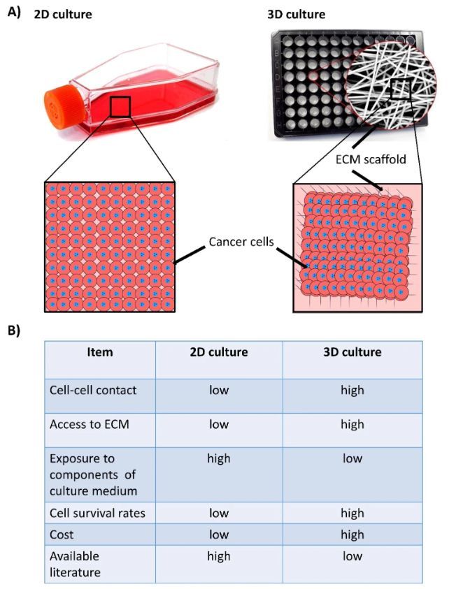 Fig.1 Major differences between two-dimensional (2D) and three-dimensional (3D) culture systems of cancer cells. (Farouk S. M., et al. 2023)
The 3D cellular scaffold model also facilitates the study of complex cellular behaviors and processes, including cell migration, proliferation, differentiation, and response to stimuli or drugs. It allows researchers to investigate tissue development, disease progression, and therapeutic interventions in a more accurate and representative manner.
Our Services
Alfa Cytology offers bladder cancer modeling services based on various material scaffolds. These scaffolds are designed to create a three-dimensional (3D) environment that mimics the tumor microenvironment and facilitates the study of bladder cancer biology, drug responses, and therapeutic interventions. Here are some of the material scaffolds utilized by Alfa Cytology in their bladder cancer modeling services:
| Type of Scaffolds |
Advantages |
Disadvantages |
 |
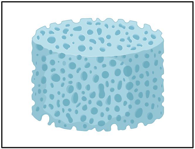
Hydrogels |
- Tissue-like responsiveness.
- Water-soluble factors are easily supplied to cells.
- Generally biocompatible.
- Low immunogenicity.
|
- Mechanical resistance is minimal.
- Physically cross-linked gels are weak.
|
 |
Decellularized scaffolds |
- Provides ECM environment.
- High bioactivity.
- Low immunogenicity.
- Promotes cell-material interactions.
|
- Decellularization of thick tissues can be difficult.
- The number of cell adhesion sites is limited.
|
 |
Fibrous scaffolds |
- Characterized by high surface-area-to-volume favoring cell proliferation, migration, adhesion and differentiation of cells
|
- Low structural stability
- Limited by cell seeding
- Scaffold morphology is difficult to regulate.
- Limited in thickness and small pore size.
|
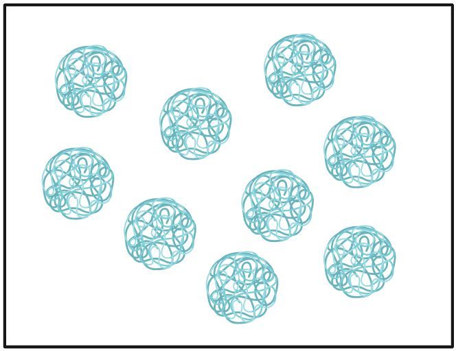 |
Microsphere scaffolds |
- Cumulative release of encapsulated bioactive substances
- Long-time maintenance of cancer cells in culture
- Excellent mechanical properties
|
- May results in loss of bioactivity of encapsulated factors.
- Residual solvent toxicity.
- Expensive.
|
 |
Nanoparticle incorporated scaffolds |
- Tunable surface properties
- High penetration ability
|
|
Case Study - The UM-UC3 Bladder Cancer 3D Scaffold Model
Model Introduction
The three-dimensional (3D) scaffold model leverages a customized bladder-mimetic hydrogel to establish a standardized, physiologically relevant in vitro platform for bladder cancer research and drug testing. It effectively recapitulates key histological features of high-grade urothelial carcinoma by culturing the human bladder cancer cell line UM-UC3 within a biomimetic extracellular matrix, providing a superior and reproducible alternative to traditional 2D cultures for preclinical therapeutic evaluation.
Model Information
- Model: UM-UC3 Bladder Cancer 3D Scaffold Model
- Cell Components: UM-UC3 Human Bladder Carcinoma Cells
Model Construction
The 3D T24 scaffold model was established according to the following standardized protocol:
- STEP 1: Scaffold Preparation – The bladder-mimetic hydrogel scaffold was prepared as 5 mm diameter discs and placed on cell culture inserts within multi-well plates.
- STEP 2: Cell Seeding & Integration – A total of 1.5 × 10⁶ UM-UC3 cells were inoculated into the scaffold at three distinct time points (day 0, 3, and 5) to ensure robust cell infiltration and proliferation.
- STEP 3: Culture Maturation – Seeded constructs were cultured for a total of 14 days under standard conditions, with medium refreshed every other day, to allow for the formation of dense, tumor-like microtissues.
- STEP 4: Drug Treatment & Analysis – For drug testing, mature microtissues were treated with therapeutic agents (e.g., Cisplatin, Gemcitabine) in multiple cycles, followed by viability assays and comprehensive histological analysis.
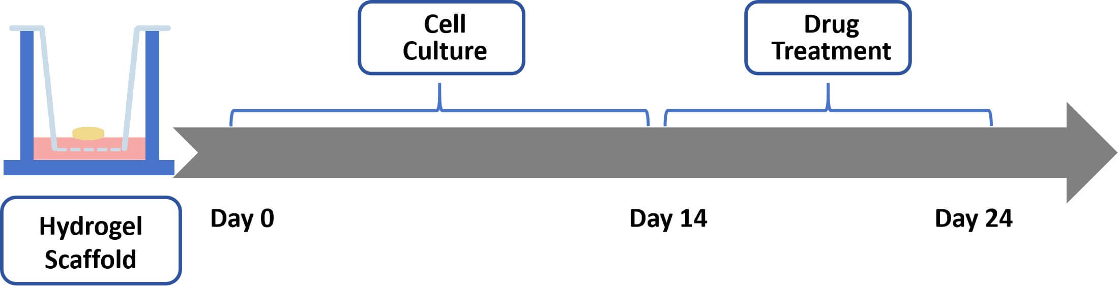 Fig. 2 Schematic workflow for establishing the UM-UC3 bladder cancer 3D scaffold model. (Source: Alfa Cytology)
Model Data
- High Histological Fidelity: The model recapitulates in vivo-like tumor architecture characteristic of high-grade urothelial carcinoma.
- Quantifiable Drug Response: Treatment with therapeutic agents induced dose-dependent and statistically significant reductions in cell viability (measured by luminescence).
- Enhanced Predictive Relevance: The 3D scaffold architecture provides a more physiologically relevant environment for drug penetration and response testing compared to 2D monolayer cultures.
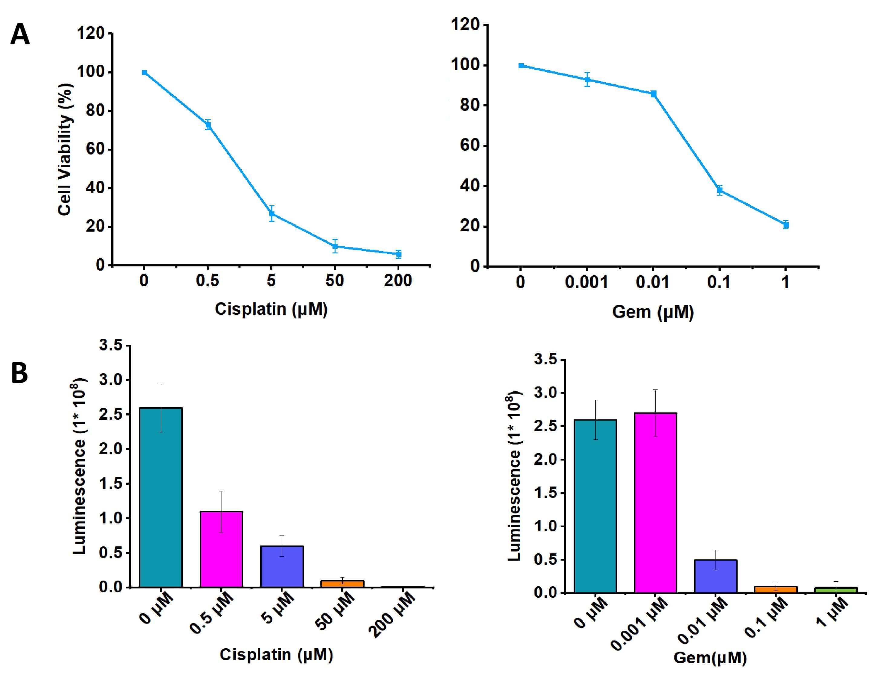 Fig. 3 Representative viability data demonstrating dose-dependent treatment response in the UM-UC3 3D scaffold model. Data are presented as mean ± SD. (Source: Alfa Cytology)
Contact Us
At Alfa Cytology, we are dedicated to providing comprehensive preclinical CRO services in the field of bladder cancer. To learn more about our scaffold-based bladder cancer modeling services or to discuss your specific research requirements, please contact us.
Reference
- Farouk S. M., Khafaga A. F., and Abdellatif A. M. Bladder cancer: therapeutic challenges and role of 3D cell culture systems in the screening of novel cancer therapeutics. Cancer Cell Int. 2023, 23, 251.
For research use only. Not intended for any clinical use.
Related Services