3D-Bioprinted Bladder Cancer-On-A-Chip Development Services
Bladder cancer-on-a-chip refers to a bioengineered in vitro model that recreates the structure and function of the bladder tissue and its associated microenvironment for the purpose of studying bladder cancer. At Alfa Cytology, we specialize in 3D-bioprinted bladder cancer-on-a-chip development.
Introduction of 3D-Bioprinted Bladder Cancer-On-A-Chip
3D bioprinting technology create a miniature, bioengineered system that mimics the complex characteristics of bladder cancer. Here's an overview of the key components of 3D-bioprinted bladder cancer-on-a-chip:
| Key Components |
Descriptions |
| Bioprinting Technology |
Bioprinting is used to create a scaffold or framework that mimics the architecture of the bladder tissue. |
| Cell Types |
The chip incorporates various cell types including bladder epithelial cells, cancer cells, stromal cells, immune cells, and endothelial cells. |
| Biomaterials |
Hydrogels such as collagen, alginate, or gelatin are commonly employed as bioinks in the bioprinting process of bladder cancer-on-a-chip models. |
| Tumor Microenvironment (TME) |
The model aims to recreate the complex TME including factors such as oxygen and nutrient gradients, extracellular matrix components, immune cell infiltration, and intercellular signaling. |
The bladder cancer-on-a-chip technology offers several advantages over traditional cell culture or animal models. It provides a more physiologically relevant platform for studying bladder cancer, allowing for improved understanding of tumor biology, drug responses, and personalized medicine approaches. Additionally, it reduces the reliance on animal models and offers a more ethical and cost-effective alternative for preclinical testing.
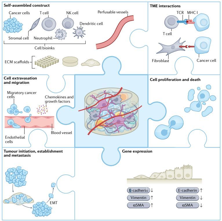 Fig.1 3D bioprinted models of cancer cell growth, migration, invasion, stemness and gene expression. (Neufeld L., et al. 2022)
Our Services
At Alfa Cytology, we offer a comprehensive range of services in the field of 3D-bioprinted bladder cancer-on-a-chip development. Our customized chips for application models provide researchers with powerful tools to study various aspects of bladder cancer biology, drug discovery, and toxicity testing. Some of the key services we offer include:
- Micro-physiology System Chips
We offer a micro-physiology system-on-a-chip that recreates the complex microenvironment of the bladder, enabling researchers to study the interactions between different types of cells, assess responses to therapeutic drugs, and gain insight into tumor development and progression.
- Drug Discovery/Screening Chips
Our microarray models are characterized by a closer fit to the physiological or pathological environment as well as high throughput, enabling our customers to assess drug responses in a faster, more accurate and predictive manner.
- Disease Modeling Chips
We offer customized microarrays that mimic specific subtypes of bladder cancer, such as non-muscle-invasive bladder cancer (NMIBC) or muscle-invasive bladder cancer (MIBC). These chips incorporate bladder cancer cells to closely mimic the characteristics of the disease, including the invasion of cancer cells into bladder tissue.
- Pharmacological Assay Chips
Our chips replicate the complex cellular and fluidic interactions that occur within the bladder microenvironment. By integrating multiple cell types and incorporating physiological flow conditions, our chips allow clients to evaluate drug absorption, distribution, metabolism, and excretion.
Case Study - 3D Bioprinted T24 Bladder Cancer Scaffold Model
Model Introduction
The model is a three-dimensional (3D) in vitro culture system for T24 bladder cancer, constructed using hydrogel as a bioink through 3D bioprinting technology. It is designed to mimic the three-dimensional architecture, extracellular matrix environment, and cell-cell interactions of in vivo tumors, providing an in vitro platform that more closely resembles physiological conditions for studying bladder cancer biology and drug responses.
Model Information
- Model: 3D Bioprinted T24 Bladder Cancer Scaffold Model
- Cell Line: T24 Cells
Model Construction
The model is established following a standardized 3D bioprinting workflow:
- Step 1: Bioink Preparation – T24 cells are uniformly mixed with a hydrogel prepolymer solution at a defined density to prepare a printable cell-laden bioink composite.
- Step 2: 3D Structure Printing – Using a temperature-controlled 3D bioprinter under optimized conditions, the bioink is precisely deposited layer-by-layer through a fine nozzle according to a predefined three-dimensional grid architecture.
- Step 3: Photocrosslinking & Solidification – Immediately after printing, the structure is irradiated with UV light at a specific wavelength to induce hydrogel crosslinking, forming a stable 3D cell-gel composite scaffold that maintains its integrity in culture medium.
- Step 4: In Vitro Culture & Experimentation – The crosslinked 3D scaffold is transferred to a culture system and maintained under standard conditions for subsequent assays, including cell viability, proliferation, and drug treatment evaluations.
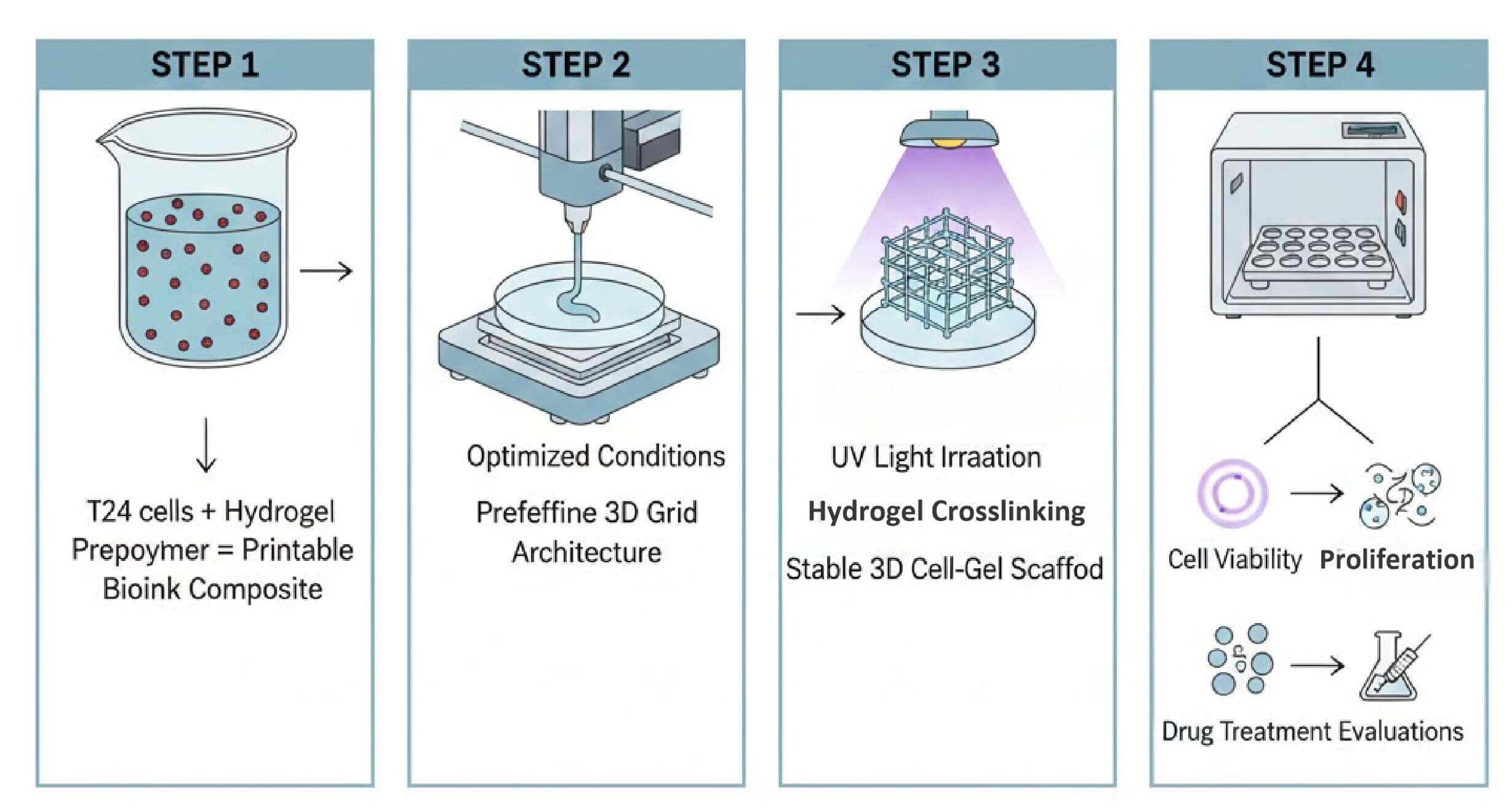 Fig. 2 Schematic workflow for establishing the 3D bioprinted T24 bladder cancer scaffold model. (Source: Alfa Cytology)
Model Data
- Enhanced 3D Proliferation: T24 cells exhibit significantly different proliferation kinetics in the 3D scaffold compared to 2D culture.
- Reduced Drug Sensitivity: Both drug A and drug B show diminished inhibitory effects on cell viability and mTOR pathway modulation in the 3D environment versus 2D.
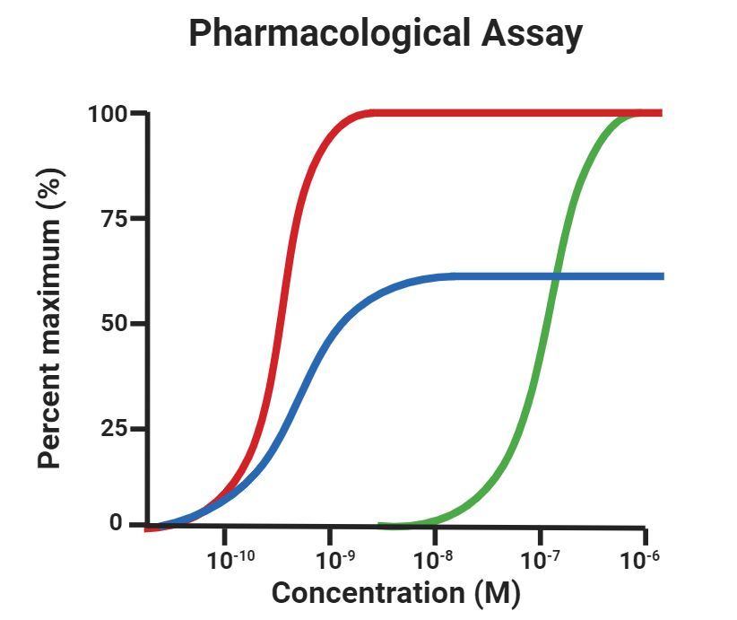
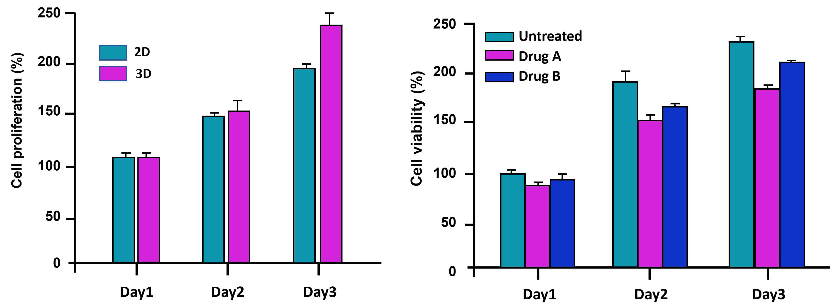 Fig. 3 Cell proliferation and drug response in 3D bioprinted bladder cancer model. Data are presented as mean ± standard error (SEM). (Source: Alfa Cytology)
Contact Us
At Alfa Cytology, we are dedicated to providing comprehensive preclinical CRO services in the field of bladder cancer. To learn more about our customized bladder cancer chip development sevices or to discuss your specific research requirements, please contact us.
Reference
- Neufeld L., Yeini E., and et al. 3D bioprinted cancer models: from basic biology to drug development. Nat Rev Cancer. 2022, 22, 679–692.
For research use only. Not intended for any clinical use.
Related Services