In Vivo Modeling Services for Bladder Cancer
In vivo models serve as powerful tools for studying bladder cancer due to their ability to recapitulate the complex tumor microenvironment, disease progression, and therapeutic responses observed in humans. At Alfa Cytology,we provide our clients with comprehensive in vivo modeling services for bladder cancer.
Introduction of In Vivo Models for Bladder Cancer
Bladder cancer is a prevalent malignancy that affects a significant number of individuals worldwide. It is crucial to develop effective therapy strategies and advance our understanding of this disease. In vivo modeling plays a critical role in bladder cancer research, allowing scientists to study the disease in a controlled environment that closely mimics the in vivo conditions.
In vivo models are shown below:
|
Types |
Description |
Syngeneic Models
- Mouse cells / tumor tissue in mice;
- Rat cells / tumor tissues in rats.
|
Subcutaneous injection / engraftment |
- Genetically engineered murine cell lines / primary cells
|
| Orthotopic engraftment |
- Primary urothelial or bladder cancer cells impaired in bladder wall of recipient host so that tumors arise in the bladder
|
| Spontaneous/Experimental metastasis |
- Spontaneous metastasis from primary site
- Intravenous (lV) injection of a bolus of tumor cells
|
Xenograft Models
- Human cells / tissues in immunocompromised mice.
|
Patients-derived tumors (PDX) |
- Potential as model individual tumors and subtypes
- Preclinical testing of specific therapeutics
- Tumor tissues are implanted in ectopic site
- Variable take rate depending on tumor subtype / phenotype
|
| Experimental metastasis |
- lV injection of a bolus of human tumor cells
- Genetically engineered human cell lines/primary cells
- Useful for studying organ tropism and later stages of metastasis
- Experimental therapeutics
|
Carcinogen-based Models
- Treatment of mice with carcinogen in drinking H2O most common is BBN.
|
- Recapitulates human disease with prolonged environmental exposure Useful (Diagnostic Biomarkers)
|
Genetically Engineered Models (GEMs)
- Expression of oncogenes and for tumor suppressors in the urothelium.
|
- Limited development of invasive and metastatic disease
- Conditional or inducible recombination of oncogenes or tumor suppressor genes in the urothelium
|
Our Services
At Alfa Cytology, we provide a wide range of in vivo modeling services tailored specifically for bladder cancer research. Our comprehensive services encompass every stage of the research process, from protocol design to project completion, ensuring that our clients receive the support they need to achieve their research goals efficiently. Here are some key aspects of our in vivo modeling services:
Case Study - UM-UC-3 Xenograft Model in C-NKG & BALB/c Mice
Model Introduction
The UM-UC-3 human bladder cancer xenograft model provides a validated preclinical platform for studying bladder carcinoma growth and therapeutic response. UM-UC-3 cells are a human bladder transitional cell carcinoma (TCC) cell line established from a primary high-grade tumor. This cell line is characterized by mutated p53 and represents a highly aggressive, basal-like molecular subtype.
Model Information
- Model: UM-UC-3 Xenograft Model
- Animals: C-NKG Mice; BALB/c Mice
- Age: 6-8 Weeks
Model Construction
The model was established by subcutaneously implanting human UM-UC-3 bladder cancer cells into mice. Tumor growth is monitored regularly by caliper measurements to track progression. Cell inoculum is 5×10⁶ cells/mouse (C-NKG) or 2×10⁶ cells/mouse (BALB/c).
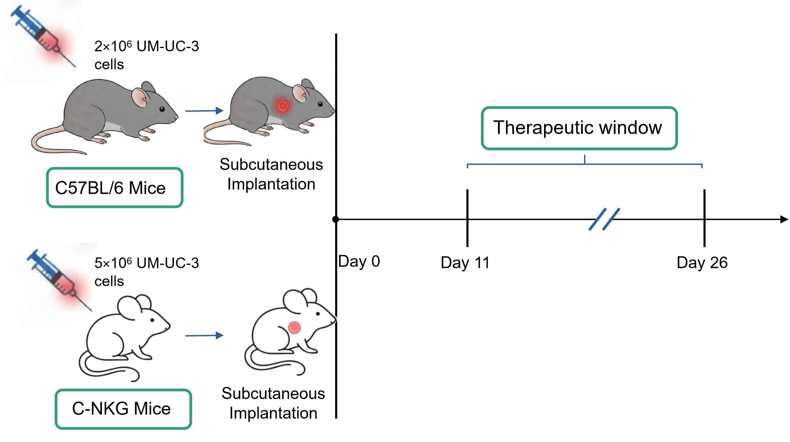 Fig. 1 Workflow of UM-UC-3 xenograft model establishment. (Source: Alfa Cytology)
Model Data
- Tumorigenicity: UM-UC-3 cells demonstrate high tumorigenic potential in both C-NKG and BALB/c mouse strains.
- Growth Kinetics: Tumor growth is rapid and consistent. Approximately 11-12 days post-inoculation, tumors reach 100-200 mm3 (suitable for initiating treatment). By around day 26, tumors approach the experimental endpoint volume of ~2000 mm3.
- Therapeutic Window: The model provides a clear treatment observation window of approximately 15 days, ideal for efficacy evaluation during the rapid tumor growth phase.
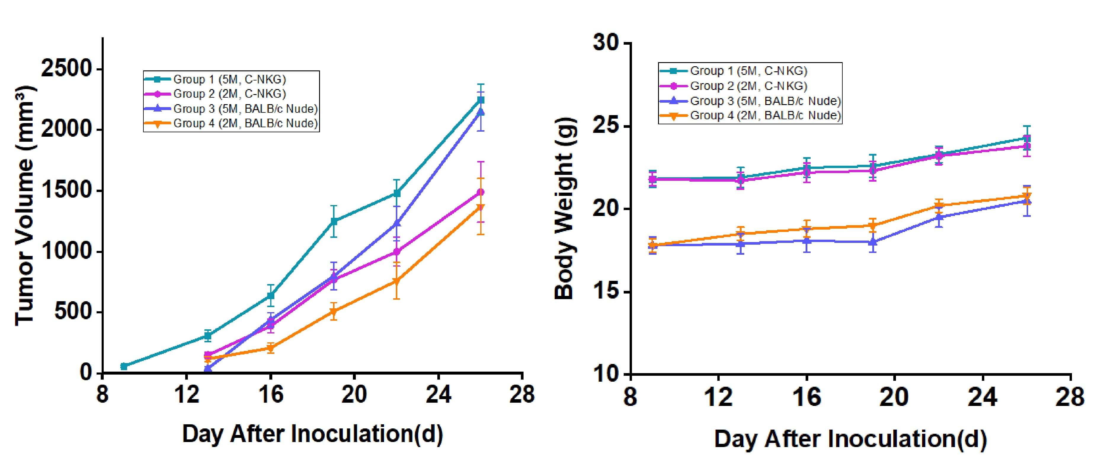 Fig. 2 Growth curves of UM-UC-3 subcutaneous xenograft tumors and mouse body weight in C-NKG and BALB/c mice (n=6). Data are expressed as mean ± SEM. (Source: Alfa Cytology)
Contact Us
At Alfa Cytology, we are committed to providing exceptional in vivo modeling services for bladder cancer research. Our experienced team of scientists, state-of-the-art facilities, and extensive range of capabilities make us a preferred partner for researchers worldwide. Whether you require the development of PDX models, GEMMs, syngeneic models, or orthotopic models, we have the expertise and resources to meet your specific research needs.
To learn more about our in vivo modeling services for bladder cancer or to discuss your research project, please contact us today.
For research use only. Not intended for any clinical use.
Related Services