Cell Line-Derived Xenograft Model Development Service
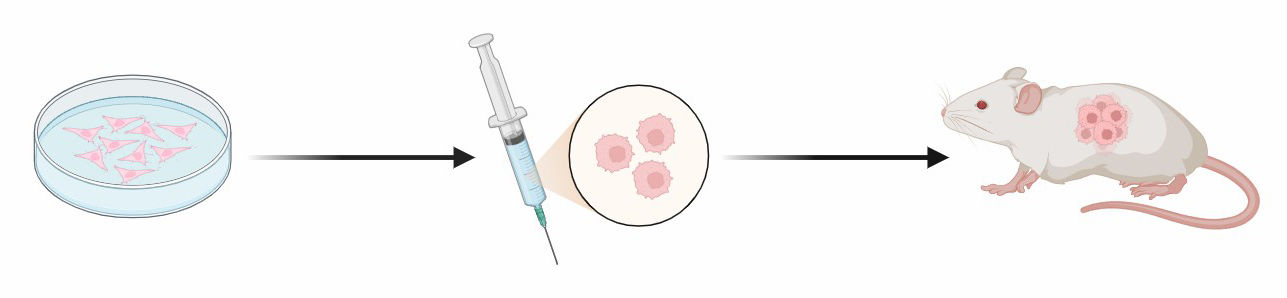
The cell line-derived xenograft tumor (CDX) model is a tumor model constructed by transplanting an in vitro cultured xenogeneic (human-derived) tumor cell line into an immunodeficient mouse. Alfa Cytology provides customized CDX models for bladder cancer research.
Cell Line-Derived Xenograft (CDX) Models
CDX models involve the transplantation of bladder cancer cell lines into immunodeficient animals, typically mice. These cell lines are derived from human and possess specific genetic and molecular characteristics. CDX models offer several advantages in preclinical research, including ease of use, reproducibility, and scalability. By utilizing CDX models, we can investigate tumor biology, evaluate therapeutic efficacy, and elucidate underlying mechanisms of bladder cancer progression.
Cell Line-Derived Xenograft (CDX) Model
Tumor Cells
Immunodeficient mouse
Transplant
They offer several advantages that enhance preclinical drug efficacy testing and analysis.
- Accessibility and Reproducibility
- Experimental Flexibility and Efficiency
- Cost-Effectiveness and Scalability
- Mechanistic Insights
Our Services
Alfa Cytology, a leading preclinical contract research organization (CRO), offers a variety of customization services for cell line-derived xenograft mouse models to develop novel bladder cancer therapies.
|
|
| Through rigorous screening and characterization, we ensure the authenticity, stability and genetic fidelity of the selected cell lines. The cell lines mainly used to construct CDX models were 5637, 5637-luc, UMUC3, 786-o-luc, RT4, SW-780. |
Taking into account factors such as implantation efficiency, tumor growth kinetics, and compatibility with the selected cell line, we provide you with the most appropriate strain, such as nude mice, NOD SCID mice, or M-NSG mice, to ensure optimal tumor engraftment and growth. |
|
|
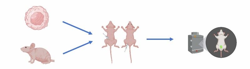
| We offer accurate and reproducible tumor cell implantation. Whether via subcutaneous or in situ (bladder), we carefully implant bladder cancer cells into mice to optimize tumor growth. |
Our regular monitoring of tumor growth using non-invasive imaging techniques such as bioluminescence or ultrasound allows for longitudinal assessment and accurate evaluation of therapeutic efficacy. |
Our Services Will Help You Achieve the Goals as Follows:
CDX models are increasingly being used to enable the following types of preclinical research for bladder cancer:
- Anti-cancer Drug Screening
- PK/PD Studies for Bladder Cancer Drugs
- Biomarker and New Drug Target Development
- Immuno-Oncology Research
- Exploring Tumor Biology and Metastasis
- Evaluation of the Efficacy of Combination Therapy
Case Study - SW780 Xenograft Model in C-NKG Mice
Model Introduction
The SW780 human bladder cancer xenograft model provides a validated preclinical platform for studying bladder carcinoma growth and therapeutic response. SW780 cells are a human bladder transitional cell carcinoma (TCC) cell line established from a primary Grade II tumor. This cell line is characterized by wild-type p53 and represents a classic luminal papillary molecular subtype.
Model Information
- Model: SW780 Xenograft Model
- Animals: C-NKG Mice
- Age: 6-8 Weeks
Model Construction
The model was established by subcutaneously implanting human SW780 bladder cancer cells into mice. Tumor growth is monitored regularly by caliper measurements to track progression. Cell inoculum is 3×10⁶ cells/mouse or 1×10⁶ cells/mouse.
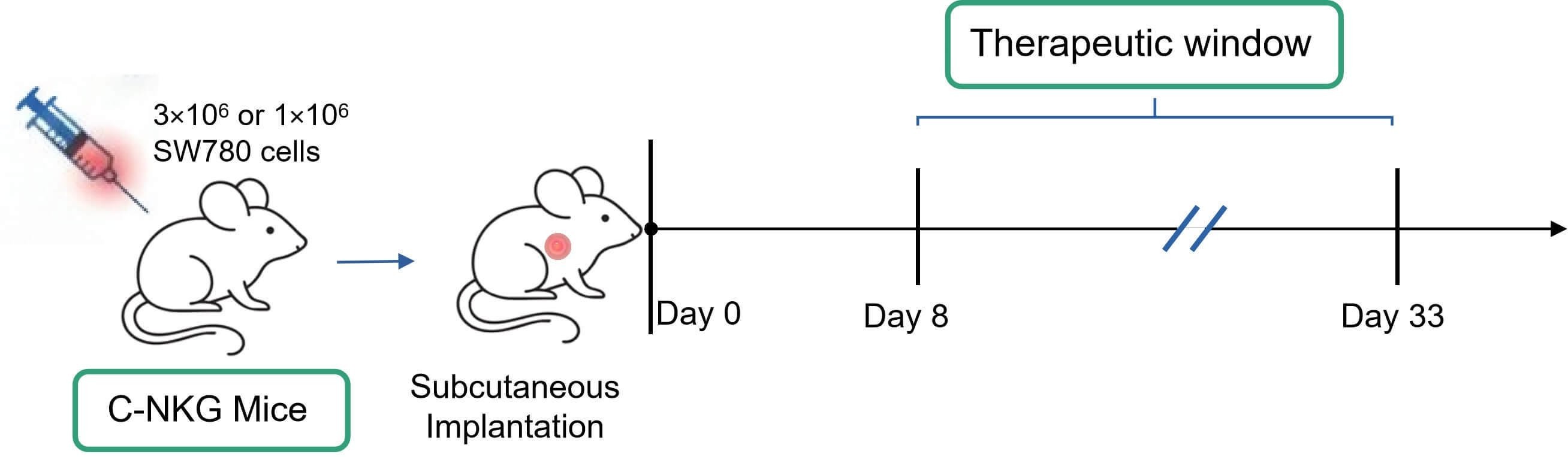 Fig. 1 Workflow of SW780 xenograft model establishment. (Source: Alfa Cytology)
Model Data
- Tumorigenicity: SW780 cells demonstrate high tumorigenic potential in C-NKG mice.
- Growth Kinetics: Tumor growth is consistent. Approximately 7-8 days post-inoculation, tumors reach 100-200 mm3 (suitable for initiating treatment). By around day 33, tumors approach the experimental endpoint volume of ~2000 mm3.
- Therapeutic Window: The model provides a clear treatment observation window of approximately 25 days, ideal for efficacy evaluation during the rapid tumor growth phase.
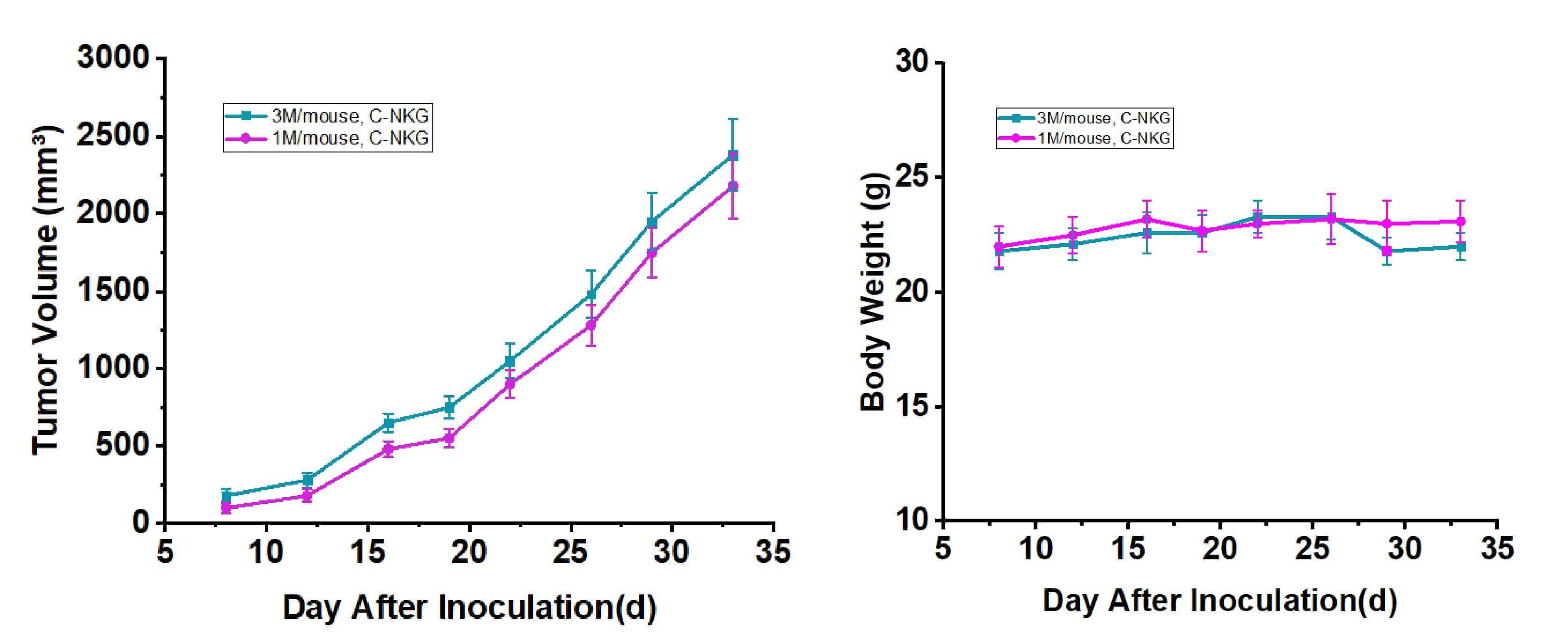 Fig. 2 Growth curves of SW780 subcutaneous xenograft tumors and mouse body weight in C-NKG mice (n=6). Data are expressed as mean ± SEM. (Source: Alfa Cytology)
Contact Us
In the quest for improved diagnostics and treatments for bladder cancer, the development of reliable and clinically relevant preclinical models is of paramount importance. Through meticulous cell line selection, precise implantation techniques, in vivo pharmacology studies, and rigorous data analysis, Alfa Cytology empowers our clients to make significant strides in understanding bladder cancer biology and identifying promising therapeutic candidates. If you have any needs, please contact us.
Reference
- Cai E. Y., Garcia J., and et al. A bladder cancer patient-derived xenograft displays aggressive growth dynamics in vivo and in organoid culture. Sci Rep. 2021, 25, 11(1): 4609.
For research use only. Not intended for any clinical use.
Related Services